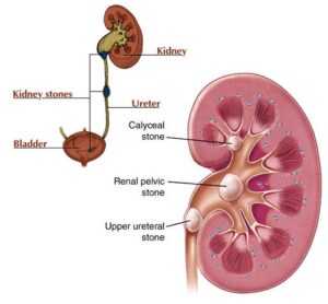
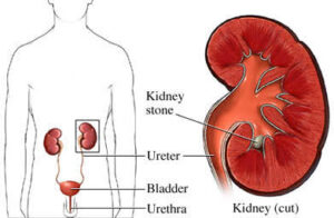
Kidney stones are the impact of the development of broke up minerals on the inward side of the kidneys. They typically comprise of calcium oxalate however might be made out of a few different mixes. The stones might be little and pass unnoticed through the urinary tract, however, they can likewise cause outrageous torment as they leave the body.
Dignosis of Kidney Stone :
Kidney Stone specialist in Dr. ANURAG GUPTA will tell you a kidney stone is a Renal calculi or kidney stones are solid pieces of crystals, salt and minerals that begin in our kidneys. However, they are likely to develop anywhere along our urinary tracts such as kidneys, ureters, bladder, and urethra. And kidney specialist give you kidney stone treatment .
Dr. Anurag Gupta is one of the Best Kidney Specialist expert in proper diagnosis and treatment of kidney stone. He is expertise in minimally invasive surgical operation and known as one of the Best Urologists. He has treated successfully different sizes of kidney stones with very good results. Dr. ANURAG GUPTA is extremely popular and Best kidney stone doctor with good results and 100% success. A highly renowned medical professional in his field of work. He follows an ethical strategy in his work and continues dedication with his patients, following the treatment method that is most proper for them. He provides the Best Kidney Stone Treatment.
Signs and Symptoms of Kidney Stone
- Pain or burning while urination.
- Pain in the backbone, stomach, belly or side.
- Blood in the urine.
- Urgent need to go.
- Going a little amount at a time.
- Dark or smelly urine.
- Fever and chills.
- Illness and vomiting.
Treatment options :
Treatment is tailored according to the types of stone. Urine can be strained and stones gathered for evaluation.
Drinking six to seven glasses of water per day increases urine flow. People who are dehydrated or have critical nausea and vomiting may require intravenous fluids.
Treatment Option Includes:
- Shockwave lithotripsy (SWL)
- Ureteroscopy and laser lithotripsy
- Percutaneous Nephrolithotomy (PCNL)
- Retrograde intrarenal surgery (RIRS)
- Lap assisted PCNL for ectopic kidneys and endoscopic surgery are some of the common surgical methods used for the effective treatment or removal of kidney stones.
Type of kidney stone:
- Calcium Stones
- Uric Acid Stones
- Struvite Stones
- Cystine Stones
1. Calcium oxalate stones
These Stones are the most common type of kidney stone. Kidney stones are hard masses that develop in the kidney when there are large levels of calcium, cysteine, oxalate, or phosphate and the too little amount of liquid. There are various types of kidney stones. Your doctor can test your stones to see what type you have. Calcium oxalate stones are created by too much oxalate in the urine.
2. Uric acid stones
These are the most common reason of radiolucent kidney stones in children. Some products of purine metabolism are almost insoluble and can precipitate when urinary pH is low. These cover 2 to 8 xanthine, dihydroxyadenine, adenine, and uric acid. The crystals of uric acid may start calcium oxalate precipitation in metastable urine concentrates.
3. Cystine kidney stones:
Cystine kidney stones are because of cystinuria, genetic disorder of the transportation of an amino acid which is a building block of a protein called cystine that occurs in an overflow of cystine in the urine and the formation of cystine stones.
4. Struvite stones
Struvite stones are a type of solid mineral deposit that can develop in your kidneys. Stones develop when minerals like calcium and phosphate crystallize inside your kidneys and hold together. Struvite is a mineral that’s created by bacteria in your urinary tract.
PCNL
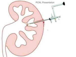
Overview
Percutaneous nephrolithotomy is a procedure used to remove kidney stones from the body when they can’t pass on their own. A scope is inserted through a small incision in your back to remove the kidney stones.
Percutaneous nephrolithotomy is used most often for larger stones or when other procedures, such as extracorporeal shock wave lithotripsy or ureteroscopy, are unsuccessful or not possible.
Why it’s done
Percutaneous nephrolithotomy is typically recommended in the following situations: Large kidney stones are blocking more than one branch of the collecting system of the kidney (known as staghorn kidney stones)
Kidney stones are larger than 0.8 inch (2 centimeters) in diameter Large stones are in the ureter Other therapies have failed Before you undergo percutaneous nephrolithotomy, your doctor will perform several tests. Urine and blood tests check for signs of infection or other problems, and a computerized tomography (CT) scan determines where the stones are in your kidney.
Risks
- The most common risks from percutaneous nephrolithotomy include the following:
- Bleeding
- Infection
- Injuries to the kidney or other organs
- Incomplete stone removal
How you prepare
Percutaneous nephrolithotomy is usually performed in the hospital under general anesthesia, meaning you will be asleep during the procedure and not feel any pain.
Your doctor may prescribe antibiotics to reduce your chance of developing an infection after the procedure.
What you can expect
Before the procedure
Before the procedure, your surgeon may have you go to the radiology department, where a radiologist will use CT, ultrasound or X-ray imaging to guide access to the kidney. This is done with a local anesthetic, meaning you will be awake but will not feel any pain. You will then be transferred to the operating room for the actual procedure.
Alternately, your surgeon may prefer to access the kidney in the operating room while you are under general anesthesia. In this case, the surgeon will put a tube through your bladder and up into your kidney, and will use X-ray imaging to make a puncture in your kidney so that the stones can be removed. If you have very large stones, the surgeon may need to make more than one puncture.
During the procedure
When the doctor has access to the kidney stones, he or she will place a sheath into the kidney and break up the stones using a special instrument or laser. When the fragments are small enough, the doctor will remove them. When the procedure is complete, the surgeon may leave drainage tubes in the kidney.
Your doctor may send the kidney stone fragments to be analyzed or to check for infection.
After the procedure
ou may stay in the hospital for one or two days after the procedure. Your doctor will recommend that you avoid heavy lifting, and pushing or pulling for two to four weeks. You may be able to return to work after a week.
If the doctor has left drainage tubes in the kidney, you’ll need to watch for any bleeding. If you notice thick, ketchup-like blood or blood clots in your urine or drainage tube, go to the emergency department.
If you develop a fever or chills, contact your doctor. These could be signs and symptoms of infection. Your doctor will probably take urine and blood tests and X-rays and then send you to the emergency department. If you have significant pain that is not relieved by your prescribed pain medicine, contact your doctor.
Results
Your doctor probably will want to see you again after four to six weeks. At that visit, your doctor may use ultrasound or X-rays to check for any stones that may be left and to make sure that urine is draining normally from the kidney.
Your doctor will also want to do some other tests to help determine what caused the kidney stones. You will also discuss ways to prevent getting more kidney stones in the future.
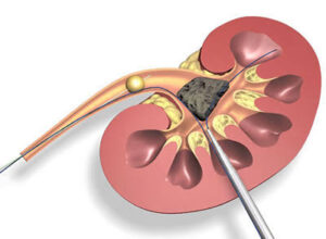
URETEROSCOPY
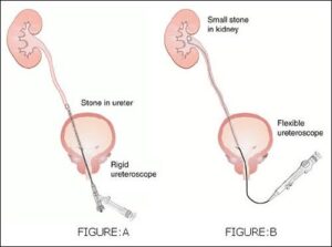
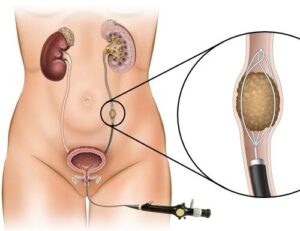
Ureteroscopy is a procedure to address kidney stones, and involves the passage of a small telescope, called a ureteroscope, through the urethra and bladder and up the ureter to the point where the stone is located. Ureteroscopy is typically performed under general anesthesia, and the procedure usually lasts from one to three hours.
If the stone is small, it may be snared with a basket device and removed whole from the ureter. If the stone is large, or if the diameter of the ureter is narrow, the stone will need to be fragmented, which is usually accomplished with a laser. Once the stone is broken into tiny pieces, these pieces are removed.
The passage of the ureteroscope may result in swelling in the ureter. Therefore, it may be necessary to temporarily leave a small tube, called a ureteral stent, inside the ureter temporarily to ensure that the kidney drains urine well.
Ureteroscopy usually can be performed as an outpatient procedure, however; patients may require an overnight hospital stay if the procedure proves lengthy or difficult.
Advantages of Ureteroscopy
Ureteroscopy can treat stones located at any position in the ureter and kidney. Additionally, ureteroscopy allows the treatment of stones that cannot be seen on an x-ray. Certain patients who cannot be treated with ESWL or PERC, such as those who cannot safely stop taking blood thinners, women who are pregnant, and the morbidly obese, can be treated by ureteroscopy.